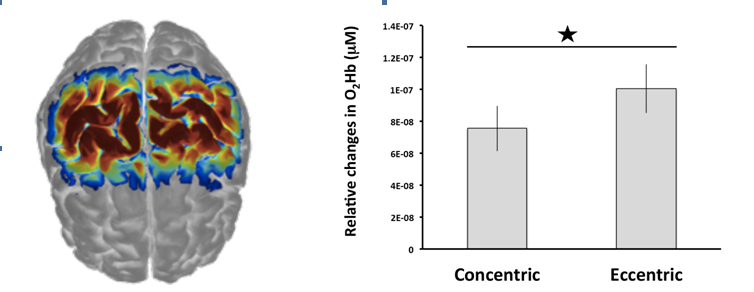
Posters● Hemodynamic response alterations in sensorimotor area as a function of muscle action: an fNIRS study The movement occurring when a muscle exerts tension while lengthening (e.g. walking down stairs) is known as eccentric muscle action. Literature contains limited evidence on how our brain controls eccentric muscle action. The latter has been shown to induce a higher cortical activation than concentric muscle actions in the contralateral sensorimotor network. However, for the primary motor cortex (M1), this observation has not reached consensus yet. It could depend on the movement velocity. Therefore, the present study aimed to investigate how M1 changes in dependency of muscle actions of the elbow flexors at two angular velocities. Nine healthy participants performed in a randomized order two exercise sessions consisting of three repetitions of five sets of maximal eccentric or concentric muscle actions at two angular velocities (30°/s and 60 °/s) for the dominant arm. The tests were carried out using a Biodex isokinetic dynamometer (System 3, Shirley, NY) with both the arm and forearm supported in the horizontal plane to avoid the effects of gravity. Dynamic muscle actions were performed over a range of motion of 60°. Hemodynamic responses based on de- and oxy-genated (O2Hb) haemoglobin for both M1s were measured with a near-infrared spectroscopy system (Oxymon MkIII, Artinis, The Netherlands) along with torque and joint angle. Intensity optical signals were first converted to optical density data before applying (i) the moving standard deviation and spline interpolation methods combined with wavelet artefact correction to remove head motion artefacts and (ii) the modified Beer-Lambert Law. Then, a band pass filter ([0.009 - 0.08 Hz]) was used to remove physiological noise.
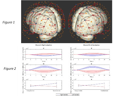
Fig.1: Left portion of the figure was generated with AtlasViewerGUI using the spatial source-detector arrangement and anatomical landmarks. Right portion of the figure shows the average (±SD) O2Hb response amplitude during eccentric and concentric tasks. *p < 0.05. Our main results (Fig. 1) showed a higher hemodynamic response (only for O2Hb, p = 0.04, partial η2 = 0.47) of the contralateral M1 to the movement during the eccentric muscle actions independently of the angular velocity. In addition, muscle actions at 30°/s have led to a significantly higher hemodynamic responses for O2Hb (p = 0.039, partial η2 = 0.48) of the contralateral M1 compared to 60°/s regardless the two muscle actions. The torque was significantly higher in concentric than eccentric muscle actions (p = 0.002, partial η2 = 0.75). Overall these findings indicate a specific control mechanism in the contralateral M1 to produce eccentric muscle actions at the movement velocities investigated. It is needed to address further this question by measuring functional coupling between M1 and other areas in the motor control network for a more comprehensive understanding of the central nervous strategies during eccentric movement as downhill walking. _______________________________________________ ● Functional brain connectivity when cooperation fails Cooperation represents a main component of our social life. In previous studies it was found that cooperative tasks are often able to improve the subjective performance and that they simultaneously contribute to modify the self-perception of social ranking position. From a neurophysiological perspective, it was found the relevant contribution of prefrontal neural areas in the case of a cooperative task specifically limbic regions and the prefrontal cortex (PFC). In addition it was found that dorsal (DLPFC) and ventral (VLPFC) portions of the lateral prefrontal cortex are generally recruited during social status inference. Studies that directly compared the effects of positive external feedback on cooperative joint actions in the case of an interpersonal performance are now numeorus. Nevertheless, at present, no specific study directly and deeply explored the influence of a negative feedback on performance and brain responsiveness simultaneously in inter-agents. Therefore, one of the aims of the present study was to investigate the effect of an external feedback (negative feedback about subjects’ performance) on the intersubjective cognitive performance and on brains’ responses. In addition, we wanted to investigate the relationship between intra and inter-brain functional connectivity by using functional Infrared-Spectroscopy (fNIRS) to address the functional connectivity effect and temporal course of the brain activation during cooperation within a hyperscanning paradigm in which participants were required to synchronize their behavioral performance. Results showed thatthe negative feedback was able to modulate participants’ responses in both behavioral and neural components. Moreover, decreased inter-brain connectivity and increased intra-brain connectivity was induced by the feedback (localized primarily in DLPFC). Finally, correlations between RTs and inter-brain connectivity revealed that negative feedback induces a more individual strategy. _______________________________________________ ● Effects of transcranial direct current stimulation (tDCS) on directed functional connectivity in the motor network derived from resting state fNIRS (Vergotte, G., Besson, P., Muthalib, M., Torre, K. & Perrey, S.) Functional near-infrared spectroscopy (fNIRS) gives a strong opportunity to combine neuroimaging and stimulation technique like transcranial direct current stimulation (tDCS). Since twenty years and the development of neuroimaging systems and analysis, the focus has changed from localization of the activation in a brain area to a more global approach based on connectivity analysis trying to understand statistical dependence between multiple areas of the brain at rest or during a task. While tDCS is a promising neuromodulation technique notably in a clinical environment, it is still unclear how tDCS could modulate functional links in the brain network under the site of stimulation and inter-hemispheric information flow. Granger Causality (GC) analyses reflect the time dependencies between two time series (or regions of interest) and thereby the directionality of the connections supposes to reflect information flow. GC analysis was used in fMRI, EEG and recently in fNIRS to investigate the connectivity between left and right hemispheres at rest. In this study, we hypothesized that tDCS at rest would alter the connectivity on the left stimulated hemisphere but in the same time could change the connectivity in the right hemisphere reflecting a reorganization of the brain to be activated in an equilibrated way. We used an fNIRS system (Octamon MkIII, Artinis Medical System, The Netherlands) composed of 2×8 channels covering the left and right sensorimotor areas (PMC and M1). The High Definition tDCS (Startim®, Neuroelectrics NE, Spain) was positioned on the left hemisphere around C3 on 9 subjects during 8-min anodal tDCS at rest after a 2-min baseline (Fig.1a). Multivariate Granger causality (MVGC) analysis was computed for all possible connections (16*16-1 channels) using the MVGC Toolbox. We then extracted the mean GC for left, right and connections between hemispheres (from left to right and right to left). Fig.1: NIRS probes and tDCS electrodes localisation (a) and Granger Causality (GC) results (mean and standard error) for the left stimulated (b) and right (c) hemispheres. a) In yellow are showed transmitters, receptors in blue, 4 return electrodes in grey and one cathodal electrode in red. Edges represent the 16 channels analysed in the study. b) and c) X-axis reflects the time and Y-axis the mean GC. Blue node = Baseline, Red = Stimulation period. Stars reflect statistical significance (p>0.05, repeated measure Anova, Bonferroni corrected). Our results highlight for the left hemisphere a progressive decrease of directed functional connectivity during tDCS (Fig. 1b). Trends for an increase of connectivity in the right hemisphere (Fig. 1c) and from the right to the left hemispheres were observed. Overall, it suggests that at resting state tDCS could cause functional reorganisation of the brain. Combining fNIRS and tDCS is promising for investigating the effects of neurorehabilitation programs in Stroke patients. Nevertheless, mechanisms involved during and after stimulation should be better investigated. _______________________________________________ ● Preparation for mental effort recruits right-lateralized Dorsolateral Prefrontal Cortex: an fNIRS investigation Preparing for a mentally demanding task calls upon cognitive and motivational resources. The underlying neural implementation of these mechanisms is receiving growing attention, given the implications for professional, social, and medical contexts. While several fMRI studies converge in assigning a crucial role to a cortico-subcortical network including Anterior Cingulate Cortex (ACC) and striatum, the involvement of Dorsolateral Prefrontal Cortex (DLPFC) during mental effort anticipation has yet to be replicated. This study was designed to target DLPFC contribution using functional Near Infrared Spectroscopy (fNIRS), as a more cost-effective tool for measuring cortical hemodynamics. We adapted a validated mental effort task, where participants performed easy and difficult mental calculation, while measuring DLPFC activity during the anticipation phase. As hypothesized, DLPFC activity increased during preparation for a hard task as compared to an easy task. Besides replicating a previous fMRI study, these results establish fNIRS as an effective tool to investigate cortical contributions to preparation for effortful behavior, especially if large samples need to be tested (to target individual differences), or populations with contraindication for undergoing fMRI (infants, elderlies or patients with metal implants), or moving subjects in more naturalistic environments (i.e. work or sport contexts). _______________________________________________ ● Neural Asymmetry for Processing Categorical and Coordinate Spatial Relations: Can fNIRS Shed a New Light on an Old Question? (Gerrits, R., Labanauskas, V., Vingerhoets, G. & Siugzdaite, R.) Introduction. Many actions involve mental representations of the way objects are located in relation to each other. Kosslyn et. al. (1987) proposed two distinct ways in which such spatial relations can be encoded. On the one hand, we can retrieve and store the precise metric distances between objects, which are referred to as coordinate spatial relations. On the other hand, we can make abstraction of the exact object position and represent its relation to other objects in categorical terms such as ‘above/below', ‘inside/outside', ‘left/right' etc. Categorical and coordinate spatial relation processing is believed to rely on distinct subsystems, which predominantly operate in the left and right hemisphere respectively. Several lesion and visual half field studies in healthy participants provide support for the suggested hemisphere asymmetry (Jager & Postma, 2003). Evidence from neuroimaging is less readily available and is equivocal (Kosslyn et al., 1998; Jager & Postma, 2003; Van der Ham et al., 2008). The present study aims to investigate categorical and coordinate spatial relations processing in healthy young adults using fNIRS. In addition, we compare left and right handers, expecting the former to show more variable/bilateral results. Methods. Twenty-eight right handers (19.6 ± 1.2 years; 14 females) and 17 left handers (average age 20.6 ± 1.8 years old; 12 females) performed a categorical and coordinate spatial relations task during fNIRS acquisition. Stimuli consisted of 2D U-shaped ‘cups', i.e. a horizontal line with a vertical line at each of its end points, as well as a single dot of 10 mm diameter, which appeared either inside or outside of the cup. During the coordinate task participants had to decide whether the vertical distance between the dot and the horizontal line of the cup was shorter or longer than the horizontal line of the cup. In contrast, during the categorical task, they had to indicate whether the dot was inside or outside the cup. Twelve 19s blocks consisting of 16 trials were presented per task condition and were alternated with rest periods of 27 to 33s. A continuous wave NIRS system with 760 and 850 nm light-emitting diodes (NIRScout, NIRx Medical Technologies, LLC) measured the hemodynamic changes. Thirty optodes (16 sources, 14 detectors) were used to cover the entire parietal lobe as well as some occipital and temporal regions, resulting in 42 channels, and a sampling rate of 3.91 Hz (see Figure 1). The fNIRS data were preprocessed using Homer2 according to the following pipeline: removal of noisy channels, normalization, wavelet based motion correction, low-pass filtering (cut-off 0.5 Hz), removal of systemic noise using the approach of Yamada et al. (2012) and GLM-modelling (gaussian basis functions). Statistical analysis consisted of mixed linear modelling (MLM) to investigate general ‘whole-template' effects of Task, Handedness and Hemisphere on ΔOxyHb, as well as a series of 2x2 ANOVA's to evaluate channel-specific effects of Task and Handedness.
Results. The MLM showed a significant Handedness by Task interaction effect (comparison to random effects only model: χ2 = 43.7, p < .001). The right handers demonstrated higher mean activation during the coordinate task, whereas the opposite was found for left handers. After applying multiple comparison correction, the two-way ANOVA procedure identified a statistically significant Handedness by Task interaction effect in channel 4 – corresponding to the right superior parietal lobe – and in channel 33, which corresponds to the left supramarginal gyrus (see Figure 1). For both channels, post-hoc t-tests show significantly higher ΔOxyHb (and lower ΔDeoxyHb) in the coordinate task compared to the categorical task in right handers. Left handers, in contrast, do not show a statistically significant difference between the two tasks. Discussion. In the present student, we aimed to investigate hemisphere asymmetry for categorical and coordinate spatial relations using fNIRS. Results show no laterality effect within the parietal lobe. First, according to the mixed linear model, only Task and Handedness effects were found to be statistically significant. Second, the 2-way ANOVA approach identified two significant channels, one in each hemisphere, that for right handers demonstrated stronger neural activity during the coordinate task. Interestingly, these results confirm those of the PET study of Kosslyn et al. (1998), where the left inferior parietal region and right superior parietal region were more strongly activated in the coordinate task compared to the categorical task. Besides replicating these findings, the present study indicates that left handers as a group do not demonstrate a clear task difference with respect to neural activity within the parietal lobe, which might suggest differential processing strategies for spatial relations between left and right handers. _______________________________________________ ● Using fNIRS in the assessment of social-communicative abilities in children from 18 to 48 months of age Joint attention is the ability to share one’s interest with others, it is a preverbal skill and one of the bases of social interaction. The cortical joint attention network is widespread and includes frontal, temporal, and parietal sites on both hemispheres. Joint attention development is deviant in individuals on the autism spectrum, and brain activation in joint attention tasks has been shown to be atypical in adults, adolescents and older children with autism spectrum disorder (ASD). Using NIRS, we have been able to get closer to the age of onset of the atypical recruitment of cortical structures during response to joint attention bids initiated by another person: for the first time, we have been testing toddlers (18 to 48 months of age) with typical development and toddlers on the autism spectrum taking part to a live joint attention task. Given the distribution of the joint attention network across several cortical areas, we used a whole-scalp probe. Preliminary results are partly consistent with those of fMRI studies investigating the joint attention network in older children and adults. Typically developing children showed a specific activation during joint attention (compared to a control attention condition) over pre-frontal, left temporal sites and the left temporoparietal junction. Children with ASD did not show a joint attention specific activation over these areas, but did show increased occipital activation during joint attention compared to the control attention condition. The recruitment of cortical areas that are proximal to those involved in the sensory processing of the presented stimuli speaks to the compromised inter-area connectivity in the autistic brain. Exploratory analysis of individual differences seems to indicate a positive correlation between activation at prefrontal sites and response to joint attention as observed during a structured play session, and between temporal activation in response to joint attention and initiation of joint attention during the play session. Prefrontal activity during response to joint attention has previously been found to correlate with better reciprocal social interaction in adults with ASD. Temporal areas are recruited both during initiation of and response to joint attention, and we can speculate that individuals with a stronger activation over temporal areas during response to joint attention can also more easily recruit these areas during joint attention initiation and thus achieve a better behavioural performance. Our findings are in line with those of previous studies over atypical brain activity during social information processing in infants who later develop ASD. Given these preliminary results, we are going to incorporate the use of fNIRS in the evaluation of an early behavioural intervention focused on the development of social communicative abilities in children with ASD. _______________________________________________ ● High density wearable-wireless diffuse optical tomography for prefrontal cortex mapping during cognitive tasks Abstract: Diffuse optical tomography (DOT) extends fNIRS by applying overlapping “high density” measurements, thus providing a 3-D imaging with an improved spatial resolution. The most recent availability of low-cost wearable continuous wave (cw) fNIRS/DOT devices is supposed to revolutionize cortical human brain mapping in the real-life [1]. The trail making test (TMT) is a widely applied diagnostic tool for measuring executive functioning. A consistent prefrontal cortex (PFC) activation was observed in young subjects while executing TMT [2]. The aim of the present study was to investigate the potential valuable use of a new high density wearable fNIRS/DOT imager for discriminating intra and extracranial oxygenation responses of the PFC in healthy subjects while executing the TMT. Methods. Eight University students performed TMT consisting of two parts (A and B). TMT-A tests the skill to connect 25 numbers consecutively; TMT-B tests the skill to connect alternatively numbers and letters (1-A-2-B-3-C..). fNIRS measurements were performed by NIRSIT (OBELAB, Republic of Korea; medical device approval in Korea – www.obelab.com) which utilizes lasers (780/850 nm) and multiple source-detector spacing (204 measurement points) to build up 3-D tomographic maps of the human cortex [3]. The 500g flexible probe covered the head in correspondence of the underlying dorsolateral and rostral PFC. Data (sampling rate 8.13 Hz) were collected by a tablet which can be located up to 7 meters from the probe worn by the subjects. For avoiding surface contaminations, the deepest investigated layer was considered for DOT data analysis (60 regions composed by 4x4 DOT voxels). Results and Conclusions. The ANOVA analysis, performed on the 60 region-DOT data, showed a significant task-related activation of the PFC. The preliminary results of this and other studies performed in our laboratory support the validity of this wearable/wireless technology to provide on-line high-density PFC activation maps. References
_______________________________________________ ● Effects of attachment styles and perspective taking on neurophysiological reactivity in young adults: a Near Infrared Spectroscopy study (Henschel, S., Doba, K., Ott, L., Pezard, L. & Nandrino, J.L.) Introduction: the quality of early child-caregiver interactions has a crucial role in the maturation of autonomic nervous system involved in stress modulation processes during socio-affective development. Disruptions in the development of a secure attachment system can alter the development of emotion regulation and empathic processes in childhood and adolescence. If many studies have highlighted the importance of attachment bonds in empathy, little is known about the neurophysiological processes involved in cognitive empathy (i.e perspective taking) in attachment disorders. Objectives: our aim was to assess the role of attachment styles on neurological patterns (Near Infrared Spectroscopy) of perspective taking during an emotional induction with attachment based pictures of three categories: distress, comfort, neutral. Pictures were presented either with the instruction to imagine the feelings of the person (‘‘3rd person’s perspective) or to imagine oneself to be in the person’s situation (“first person’s perspective”). Method: a sample of 92 participants (47 males, 45 females) with a mean age of 19,6 years old, from a general population was recruited and filled self-reported questionnaires assessing attachment, empathy, and emotion regulation. Two groups of attachment were created: secure and insecure. Changes in oxygenated hemoglobin (oxy-Hb) were examined by Near-Infrared spectroscopy system, as an indicator of regional cerebral blood flow changes, which reflects brain activity directly related to emotions. Oxy-Hb was recording during exposure to emotional pictures. Prefrontal and frontal areas were recorded, and NIRS analyses were conducted during resting, induction and recovery periods. Hypotheses: attachment styles would have differential effects during perspective taking conditions. More specifically, participants of the insecure-group would report fewer frontal levels of oxy-Hb during the first person perspective taking condition. |


