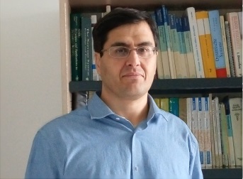
Conférenciers Invités
Groupe de recherche sur l’analyse multimodale de la fonction cérébrale, INSERM U1105, Université de Picardie Jules Verne, France.
Title : Confounding factors in event-related fNIRS analysis Abstract:In many experiments, slow/rapid event-related designs are used to temporally characterize the brain hemoynamic response (HDR) to discrete (cognitive) events. The general linear model and conventional averaging are commonly used to estimate the HDR profile in event-related designs. The accuracy of these techniques is compromised by several confounding factors. In this talk, I will discuss the effect of the main confounding factors including timing parameters of event sequences, different types of noise, signal-to-noise ratio, temporal autocorrelation and temporal filtering on the performance of these techniques in slow and rapid event-related designs.
**************************************
Dr. Robert J Cooper Department of Medical Physics and Bioengineering, University College London, UK.
Abstract: Functional near-infrared spectroscopy is a rapidly growing research field, and fNIRS technologies are an increasingly common sight in both neuroscience laboratories and in clinical environments. Diffuse optical tomography (DOT) is an extension of NIRS that uses a large number of overlapping measurements to yield three-dimensional, depth-resolved images of concentration changes in oxygenated and deoxygenated haemoglobin. However, the necessity of using optical fibre bundles continues to severely limit how far we have been able to push DOT technologies, which in turn has prevented their uptake by psychologists, neuroscientists and clinicians. In this talk, I will discuss how the advent of wearable, high-density DOT devices has already begun to revolutionize the fNIRS field, and explain why these technologies will allow more researchers to move their research out of the MRI scanner suite and into every-day environments.
**************************************
Department of Life, Health and Environmental Sciences, University of L'Aquila, Italy.
Title: The first 40 years of non-invasive medical near-infrared spectroscopy (NIRS) research: from localized brain/muscle oximetry to wearable-wireless functional NIRS/DOT Abstract: Starting with the pioneering work of Jobsis at Duke University in the 1977 [1], non-invasive near-infrared spectroscopy (NIRS) was utilized for investigating firstly cerebral oxygenation either experimentally or clinically, and later local muscle oxidative metabolism at rest and during even dynamic exercise. Since the end of the nineties of the last century several commercial two-channel brain oximeters have been available for monitoring adults and newborns with risk of brain hypoxia/ischemia. In the 1993, four research groups provided evidences of the potentialities of NIRS to assess brain activation through the intact skull in adults and also in newborns (in the 1998) [2]. It is well known that in healthy subjects neuronal activation evokes a regional cerebral blood flow increase. Therefore, the typical oxygenation response over an activated cortical area is represented by a localized increase in oxyhemoglobin (O2Hb) and a decrease in deoxyhemoglobin (HHb). This discovery has added a new dimension to NIRS research. In the middle of the nineties, the introduction of multi-channel NIRS systems, utilizing arrays of multiple near-infrared sources and detectors arranged over the scalp, led to the development of NIRS as a functional neuroimaging methodology named functional NIRS (fNIRS) [3]. More recently, diffuse optical tomography (DOT) extended fNIRS by applying overlapping “high density” measurements, thus providing a 2-D and 3-D imaging with an improved spatial resolution [4]. The recent availability of low-cost wearable continuous wave fNIRS/DOT devices is supposed to revolutionize cortical human brain mapping in the real-life [5]. Although in the last decade European researchers, including the French ones, gave a consistent contribution to fNIRS research, several technical problems are still open and they will be discussed based on the long lasting experience of our fNIRS laboratory.
|


