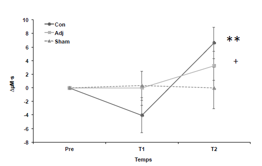
Communications orales● Evolutionary approach to emotion using affective prosody: a fNIRS study (Debracque, C.) In recent years, the emergence of affective sciences has allowed understanding how emotions are decoded by the human brain. For instance, several studies have investigated the ability to decode the speaker’s emotional state through functional neuroimaging, using affective prosody, the variations of the vocal tone in voice, as a clue. Beyond the involvement of the auditory temporal areas, these studies have suggested the role of both the right and left inferior frontal cortex (IFC) in attentive decoding and cognitive evaluation of emotional cues in human vocalizations. Hence, the bilateral IFC activation might depend on the nature of emotional vocalizations (emotional prosody versus nonverbal expressions) and on the level of attentive processing (explicit versus implicit). From an evolutionary point of view, humans are primates, belonging to the hominid family together with other great apes (chimpanzees, bonobos, orangutans, gorillas). Because of this close genetic proximity, it is likely that humans would be able to recognize expressed emotions from other apes’ vocalizations, possibly displaying variation of activation in the bilateral IFC according to the phylogenetic tree. In this study, our goal was to investigate via functional Near Infrared Spectroscopy (fNIRS, Artinis) how humans categorize and discriminate emotions in primate vocalizations within implicit or explicit modalities. All participants were exposed to the same stimuli, consisting for human voices of onomatopoeias (extracted from the Montreal Affective Voices-MAV) and calls produced by chimpanzees, bonobos and macaques for other primate vocalizations. These stimuli were expressed by two male and two female speakers in an angry, fearful or happiness tone for human voices. Equivalent calls for primate vocalizations expressed: anger (aggressors screams), fear (victim screams) and happiness (food grunts). 36 different stimuli were presented during a mini-block design with two tasks: “emotion categorization” and “emotion discrimination” repeated three times resulting in 6 blocks, the order of which was pseudo-randomly assigned to each participant. The same stimuli were used in a discrimination and categorization task to test the hypothesis of lateralization of the cerebral processing. We predicted that the emotion task would induce an increase of the OxyHemoglobin (the most relevant signal from the Hemodynamic Response function) in the Bilateral IFC more or less important and specific according to the species that produced the vocalizations (Human > Chimpanzee = Bonobo>Macaque). Furthermore, we expected that the behavioral results: time reactions (RT) and the number of correct answers would follow the same phylogenetic pattern, as the fNIRS data, with an increase of RT and a decrease of answer accuracy for macaques in opposite to chimpanzees and bonobos. We present here the preliminary results of this experiment in fNIRS. _______________________________________________ ● Sensorimotor adaptation: bridging the gap between brain networks dynamics and behavioral variability (Vergotte, G.) Number of experimental neuroimaging studies in the recent years has contributed to highlight the huge ability of the brain to exploit its inherent plasticity to adapt to various intrinsic or extrinsic constraints, over different time scales. For a sensorimotor task, the adaptation of the brain could be defined by the capacity to perform the task at the same level of performance whatever constraints. Using a simple sensorimotor paradigm as a finger-tapping task of 6 minutes and 40 seconds, we hypothesized that the deprivation of sensory feedback (Auditory and / or Visual and / or Tactile) should not change the behavior-related performance variables like the mean, the coefficient of variation and the drift. On the other hand, brain networks should evolve in responses to the deprivation of feedback. Indeed, the number of networks involved during the prolonged motor task may increase with the number of deprived feedback. Using two fNIRS systems (Octamon and Oxymon MkIII, Artinis Medical System, The Netherlands) 24 channels allowed to delineate 4 regions of interest (PFC, SMA, PMC and M1) on both hemispheres in 32 healthy subjects. We used a standard pre-processing to remove artifacts' and to extract the frequency band of interest [0.009 - 0.08 Hz]. Then a simple bivariate functional connectivity analysis (sliding window Pearson’s correlation) was applied in combination with a modularity analysis to extract the time evolution of networks during 6 minutes of the motor task. Behavioral performance did not show any differences between the four groups. On the other hand, we detected 8 distinct networks for the control group (without feedback deprivation), 16, 13 and 13 networks for groups -1, -2 and -3 feedback, respectively (Fig. 1). Fig. 1: Number of networks involved during the task for the four experimental groups, with the weight for each network and distances between them. Nodes (C) reflect number of networks extracted from functional connectivity and modularity analysis. The size of the nodes reflects the weight of each network or, in other words, its time of occurrence during the task (from 100 to 800 of 3600 data points). Links (in grey) show the Euclidean distances between networks: the more two networks are different, the greater the distance is. Our results indicated clear dynamical changes during the motor task. While a lot of studies consider that activation areas in the brain are localized and stable during a task, we highlight the dynamical organization that is a principal property of complex adaptive systems. To perform the task at the same level of performance without constraints, the brain should exploit other networks configuration. Studying such time varying organization both at the level of the brain and the behavior makes it possible to go along with conventional analyses for a better understanding of the adaptation at the cortical level using fNIRS. Our study opens new way to understand brain-behavioral link and motor adaptation. _______________________________________________ ● When cooperation goes wrong : brain and behavioral correlates of ineffective joint strategies (Gatti, L.) Cooperation, defined as a set of interactions with others that increase shared performance, is one of the most important human social behaviors. Cooperation secures a benefit to all the people engaged as well as important behaviors like acting prosocially. But what happens when the joint actions are not effective? This study aims to investigate the neural correlates of cooperation during a joint task. To do this an hyperscanning paradigm has been used which consists in the simultaneous recording of the cerebral activity of two or more subjects involved in interactive tasks. We asked 24 participants paired in 12 dyads to cooperate during an attentional task in a way to synchronize their responses and obtain better outcomes. The task was sub-divided in 8 blocks with a pause halfway assessing the goodness of the cooperation scores. The feedback was defined a priori in order to provide a social manipulation about the performance and modify their responses. The feedback was negative most of the times in order to frustrate the subjects and induce them to improve the performance in the next step. The effects of the feedback were explored by means of functional near-infrared spectroscopy (fNIRS). Results showed a specific pattern of brain activation involving the dorsolateral prefrontal cortex (DLPFC) and the superior frontal gyrus (SFG). The DLPFC showed increased O2Hb (oxy-hemoglobin) level after the feedback, compatible with the need for higher cognitive effort. Also, the representation of negative emotions in response to failing interactions was signaled by a right-lateralized effect. For the STG, instead, a decreased activity was found after the feedback, which could be interpreted as disengagement for goal-oriented social mechanisms elicited by the negative and frustrating evaluation. Results were interpreted at light of available knowledge on perceived self-efficacy and the implementation of common goals and strategies. _______________________________________________ ● Delayed increase in sensorimotor cortex activity induced by coupling motor task – anodal transcranial direct current stimulation (Besson, P.) Neuromodulation provided by transcranial direct current stimulation (tDCS) offers interesting possibilities in neuroergonomics. There is, however, some interindividual variability in cerebral responses to the anodal tDCS [1]. Many parameters have to be taken into account to optimize the effectiveness of the intervention protocol by tDCS. Coupling the tDCS when performing a motor task appears to be a promising protocol for increasing the level of performance and motor learning compared to a tDCS protocol followed by the motor task [2]. The neuroplasticity of the sensorimotor cortex (SMC) should be further accentuated. Nevertheless, no study has isolated the effects of these two types of protocol for a given motor task. The objective of this randomized controlled study was to compare with SIR spectroscopy (fNIRS) SMC activation as a function of tDCS protocol type. The hypothesis was that the tDCS anodal and motor task coupling protocol would modulate the activation of the ispilateral SMC to the tDCS in proportions greater than the tDCS protocol followed by a motor task. Nine right-handed healthy participants performed a rhythmic finger opposition task at 3 distinct times (pre: basal state, T1: during or immediately after tDCS and T2: 30 minutes after tDCS) during 3 sessions spaced a week apart interval. Each subject performed the following 3 tDCS anodal protocols (20 min to 2 mA): concurrent (i.e., tDCS during task) or adjacent (i.e., tDCS before task) or sham protocol (i.e., placebo). The anodal tDCS electrodes (configuration 4x1, [3]) were positioned on the left hemisphere at the SMC contralateral to the movement produced. SMC activation levels reflected by a cerebral oxygenation index (Hbdiff) were measured during the task via an NIRS system with 4 channels around the anode stimulation site. The results show that the participants maintained the same motor rhythm (~2.4 Hz) for the 3 protocols. At the T2 period, SMC activation was significantly higher for concurrent compared to the other 2 protocols (i.e., adjacent and sham). These results suggest that motor task coupling with anodal tDCS is more favorable to induce changes in cerebral activity after a short post-tDCS period, demonstrating neuroplastic mechanisms that alterthe excitability / inhibition equilibrium. Fig. 1. Mean (± SEM) of the relative changes (relative to the pre-period) of the Hbdiff concentration of the left sensorimotor cortex during the motor task for the 3 anodal tDCS protocols: concurrent, adjacent and sham at the 3 measurement periods: pre, T1 et T2. ** P < 0,001 et + P = 0,052 versus sham. References 1. Chew TA et al. (2015) Inter- and intra-individual variability in response to anodal tDCS at varying current densities. Brain Stimul, 8:374. _______________________________________________ ● Interpersonal Brain Synchronization Tracks Social Interactive Learning (Yafeng, P.) Throughout evolution, humans have adapted to learn from others through social interaction. However, the mechanistic features of such social interactive learning are not well understood. In this study, we examined the quantity and dynamics features of social interactive learning by the functional near-infrared spectroscopy (fNIRS) – based hyperscanning technique. Twenty-four learner-instructor pairs participated in a song interactive learning task while their brain activity was simultaneously recorded. We experimentally manipulated the amount of interactions by using two distinctive learning approaches: part (more interactions) and whole (fewer interactions). We found better learning performance and higher interpersonal brain synchronization (IBS) in the part learning (vs. whole learning), indicating contribution of amount of interactions to the performance and IBS. Learner’s performance co-varied with the detected IBS during vocal interactions, especially with those occurring at learner observation. Granger causality analyses further found the stronger directional IBS from the instructor to learner than from the learner to instructor. These effects were absent either in learner imitation or at any process in the whole learning. These results confirmed the roles of the quantity and dynamic process of social interactions in learning from others, and suggested the learner-instructor brain-to-brain communication during effective social interactive learning. Keywords: social interactive learning, interpersonal brain synchronization, part learning, whole learning, fNIRS _______________________________________________ ● Identification of the metabolic correlates of the activation/inhibition pattern: a study combining fNIRS and EEG methods (Roger, C.) The ability to choose an appropriate action under time pressure is an important feature of cognitive control. Neurophysiological evidence indicates that neural inhibition is an important component of motor processing in choice reaction time (RT) tasks. Thanks to Laplacian transformation on monopolar EEG data, Vidal et al [1] revealed two noticeable features of the motor command in between-hand choice RT tasks, called the activation/inhibition pattern. First, just before the response, a negativity develops over the contralateral primary motor cortex (M1) revealing an activation of the correct response. Second, a positivity over the ipsilateral M1 develops symmetrically, and reveals an active inhibition of the incorrect response. This activation/inhibition pattern has also been observed using transcranial magnetic stimulation and H reflex [2]. However, this specific pattern, in particular the presence of the ipsilateral inhibition, has not been observed using metabolic neuroimaging methods. The aim of the present study was to fill this particular gap using the functional Near-Infrared Spectroscopy (fNIRS) with an event related design in order to observe the metabolic correlate of the activation/inhibition pattern. For that purpose, participants performed a RT task in which they had to response as a function of the direction of arrows presented on a visual display. For some participants, fNIRS recordings were combined with EEG in order to have a control of actual presence the activities of interest. Preliminary results confirmed the classical activation/inhibition pattern in EEG data. However, the hemodynamic response appearing in the fNIRS data is identical in the left and right hemisphere. Acknowledgments This work was supported by grants from ANR-11-EQPX-0023, FEDER Sciences et Cultures du Visuel, Université de Lille, MESHS. References[1] Vidal, F et al. (2003) The nature of unilateral motor commands in between-hands choice tasks as revealed by surface Laplacian estimation. Psychophysiology, 40:796-805. |


